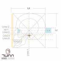
Ficha técnica I-a. Craneo / Technical data I-a. Skull.
Ficha técnica I-a.
BASES ACADEMICAS
PARA LA CONSTRUCCIÓN
DE UNA CRÁNEO HUMANO
Technical data I-a.
ACADEMIC BASIS FOR A HUMAN SKULL CONSTRUCTION
punto de partida
para la
realización de un busto
Queremos
empezar con la enseñanza de la construcción del cráneo humano en primer lugar
porque es esta parte del cuerpo humano la que va a regir como unidad de
medida para las demás partes del cuerpo y sus proporciones (normalmente la
gran mayoría de academias y centros de educación de Bellas Artes lo trabajan
de esta manera), en segundo lugar porque nos va a permitir acercarnos a la
construcción objetual de una manera muy sencilla y práctica ya que por su
figura espacial cerrada se vuelve un ejercicio muy sencillo y de fácil
realización.
|
Starting
point
for
the realization of a bust
-
We begin with the teaching of the
human skull construction first because it is this part of the human body that
will govern the unit of measure for the other parts of the body and its
proportions (usually the vast majority of Fine Arts schools and education centers
works it this way), secondly because it will allow us to get close to the objectual
construction in a very simple and practical way just because of its close spatial figure
becomes a very simple exercise and easy to make.
|
AREA
TEORICA
Anatomia
biológica.
A
continuación encontrarán ocho diferentes esquemas anatómicos que nos servirán
para reconocer y entender la estructura, ubicación y disposición de las
diferentes partes del cráneo humano. Dichas laminas muestran el cráneo desde su vista
frontal, lateral, superior, inferior, así como cuatro acercamientos y
detalles del mismo. Queremos dejar en claro que no es nuestra pretensión realizar un estudio pormenorizado, sino que
queremos únicamente hacer hincapié en este material como elemento de apoyo
objetivo y racional.
|
THEORETICAL AREA
Biological anatomy.
Below you will find eight
different anatomical diagrams that will help us recognize and understand the
structure, location and layout of the different parts of the human skull.
These sheets show the skull from the front view, side, top, bottom, and four
approaches and details. We want to make clear that it is not our intention to
carry out a detailed study, but only want to emphasize this material as an
objective and rational support.
|

COMPOSICIÓN Y PROPORCIONES ANATOMICAS / COMPOSITION AND ANATOMICAL PROPORTIONS
“El
cráneo humano tiene la medida perfecta”. No hay mayor y mas cierta expresión
que lo dicho por Vitrubio en lo que se refiere a las medidas y proporciones
del cuerpo humano. Fácilmente se darán cuenta a través de las siguientes
imágenes lo perfecto y armónico que es el cráneo humano, y la simplicidad con
que uno puede llegar no tan solo a entenderlo sino a representarlo solo con
seguir dichos esquemas como si fuesen una carta de navegación.
|
"The human skull is the
perfect fit." There is no greater and more certain expression that told
by Vitruvius in regards to the measurements and proportions of human body.
Easily will realize through the following images is perfect and harmonious
human skull, and the simplicity with which one can get not only to understand
but to continue to represent only those schemes as if they were a
navigational chart.
|
VISTA
LATERAL / SIDE VIEW
|
|
|
|
Imagen
1
|
Imagen
2
|
Imagen
3
|
Construya
o dibuje un cuadrado perfecto, si es posible intente que sus lados sean de 15
cm, como se señala en la imagen 1. Separe una sección que mida el 5 % del total del ancho como se
muestra en la imagen 2. Posteriormente divida el cuadrado de forma vertical
en dos secciones desiguales con la proporción armónica, es decir que la
sección mayor se encuentre en la parte superior y le corresponda el 0,6182
con respecto al total de la medida de la cara lateral del cuadrado (9,273 cm)
y la sección menor en la parte
inferior de 0,3818 (5,727 cm.) como se muestra en la imagen 3. De esta manera
obtendremos una línea horizontal guía A
que nos marcara la altura en la que se encuentra el orificio auditivo.
|
Construct or draw a perfect
square, if possible, try to be 15 cm its sides, as indicated in Imagen 1. Separate
a section measuring about 5% of total width as shown in Imagen 2.
Subsequently divide the square vertically into two unequal sections with golden
proportion, so the higher section of the left side has a width of 0.6182 with
respect to the total measure of the square face (9,273 cm) and lower section
with a width of 0.3818 (5,727 cm.) as shown in Imagen 3. In this way we
obtain a horizontal guide line A which
shows the height of ear orifice.
|
|
| ||
Imagen
4
|
Imagen
5
|
Imagen
6
|
Dibuje
dos líneas diagonales que unan los vértices opuestos del rectángulo restante
descontando la zona del 5% que se delimito en la imagen 2 y trace una línea
vertical y otra horizontal exactamente en la intersección de dichas líneas
oblicuas obteniendo los centros o ejes del rectángulo (Imagen 4). La línea
eje vertical en su cruce con la línea A
corresponderá al centro del orificio auditivo, mientras que la línea eje
horizontal corresponde al eje de la cavidad ocular (imagen 5). Divida ahora
la sección menor que se obtuvo al trazar la línea A en dos partes desiguales con una proporción armónica y obtendrá
dos secciones, la menor en su parte superior
(2,18656) y una sección mayor en la parte inferior (3,5404), trace una línea guía B que divida dichas secciones, dicha
línea marcará el piso del cráneo en su parte posterior (hueso occipital), así
como también el nacimiento de los incisivos del maxilar (imagen 6).
|
Draw two diagonal lines joining
opposite corners of the remaining rectangle subtracting the 5% delimited area in Imagen 2 and draw a
vertical line and another horizontal
exactly at the intersection of these oblique lines obtaining the rectangle centers
or axes (Imagen 4). The vertical axis line at its intersection with line A will correspond to the center of ear
hole, while horizontal line axis corresponds to the eye cavity axis (Imagen
5). Now divide lower section obtained
to draw the line A into two
unequal parts with a harmonic proportion and get two sections, the smallest
at the top (2.18656) and a larger section at the bottom (3.5404) , draw a
guide line B that divides these
sections, that line will mark the skull floor (base) on the back (occipital bone), as well as the
birth of the maxillary incisors (Imagen 6).
|
|
|
| |
Imagen
7
|
Imagen
8
|
Imagen
9
|
Si
usted divide la distancia que hay entre la línea eje h y la línea A en dos
partes desiguales con una proporción armónica, dejando la sección mayor en la
parte superior y la menor en la parte inferior obtendrá una línea guía C (imagen 7) que corresponderá a la
base del hueco ocular. Divida la distancia que existe entre la parte superior
de su rectángulo contenedor y la línea A
en cuatro partes iguales (imagen8). Utilice uno de los cuartos que obtuvo en
el paso anterior como medida, colocando su base sobre la línea C trazando en su parte superior una
línea horizontal C´, que marcará
el límite superior del hueco ocular (imagen 9).
|
If you divide the distance
between the axis line h and line A
into two unequal parts with a harmonic proportion, leaving the larger section
at the top and the least at the bottom get a guideline C (Imagen 7) corresponding to ocular hollow core. Divide the
distance between the top of your container rectangle and line A in four equal parts (imagen8). Use
one of the quarters obtained in previous step as measure, by placing the base
on the C line drawing a horizontal
line C’ at the top, which marks the upper limit of the eye hole
(Imagen 9).
|
|
|
|
|
Imagen
10
|
Imagen
11
|
Imagen
12
|
Divida
en partes desiguales, de forma que obtenga una proporción armónica entre ella,
la distancia que hay entre el eje vertical de su rectángulo contenedor y su
cara izquierda (acuérdese de no tomar en cuenta el área del 5% que separó en la imagen 2), obteniendo una
sección mayor en su lado derecho y una
sección menor en la parte izquierda. Trace una línea vertical 2 partiendo de
la división de las secciones antes mencionadas, dicha línea determinará la
posición más profunda del límite lateral del hueco ocular en la apófisis frontal del hueso cigomático
(imagen 10). Divida en dos secciones
desiguales en una proporción armónica la distancia entre el extremo izquierdo
de la sección que se separó en la Imagen 2 hasta la línea 2, dejando la
sección mayor del lado izquierdo y la menor del lado derecho, la línea
vertical 3´que las divide marcará el límite superior del hueco ocular.
Realice nuevamente la misma operación pero tomando la distancia solamente
desde la cara izquierda del rectángulo contenedor hasta la línea 2, la línea vertical 3 que divide esta dos secciones
mas pequeñas determinará el límite inferior de la cuenca ocular (imagen 11).
Divida en tres secciones iguales la altura del rectángulo contenedor, trace una
línea horizontal D que parta de la
división del primer tercio con el segundo tercio, dicha línea nos mostrará el
límite inferior del hueco nasal (imagen 12).
|
Divide into unequal parts, in a
way you get a harmonic proportion itself, the distance from the vertical axis
of the container rectangle and its left side (remember not to consider the 5%
area separated in image 2), obtaining a larger section on the right side and
a smaller section on the left. Draw a vertical line 2 starting from the
division of sections above, the line will determine the deepest position of
side boundary of hole ocular in frontal apophyses of zygomatic bone (Imagen 10). Divide into two unequal
sections in a harmonic proportion the distance between the left end of the
separated section in Imagen 2 to line 2, leaving the larger section in left side and the smallest in right side, the vertical line 3' that divides
them will show the upper limit of the eye hole. Make the same operation again but only by taking the distance
from the left side of container rectangle to line 2, the vertical line 3 that
divides this two smaller sections will determine the lower limit of the eye
socket (Imagen 11). Divide into three equal sections the of height container
rectangle, draw a horizontal line D that starts in the division of the first
third with the second third, the line will show the lower limit of the nasal hole
(Image 12).
|
|
| ||
Imagen 13
|
Imagen 14
|
Imagen 15
|
Multiplique
por su crecimiento armónico (1,6182) la distancia que existe entre las líneas
horizontales C y D, obteniendo una distancia tal que
realizando en su extremo inferior una línea horizontal E obtendremos la medida inferior del hueso cigomático en su
apófisis maxilar (imagen 13). Partiendo del extremo inferior de su rectángulo
contenedor trace una línea horizontal
F a una altura del 10% del total de la altura de dicho rectángulo, la
línea F marcara la altura en que
se encuentra el ángulo posterior del hueso de la mandíbula. Para obtener la
profundidad del ángulo de la mandíbula, divida en dos partes desiguales en
una proporción armónica la distancia entre las líneas verticales 2 y 1
dejando la sección mayor en el extremo izquierdo y la menor en el extremo
derecho la línea vertical 4 que las divide en su intersección con la línea F marcara el punto exacto del ángulo
de la mandíbula (imagen 14). Por último
tomando en cuenta la medida de ½
de la altura total del cráneo, partiendo desde el hueco auditivo en
forma vertical ascendente, el extremo superior de dicha medida determinará el
límite superior de la depresión lateral que tienen los huesos frontal,
temporal, esfenoides y parietal por la superposición que tendrá el musculo
temporal (imagen15).
|
Multiply by harmonic growth
(1.6182) the distance between the horizontal lines C and D, obtaining
such a distance that making a horizontal line E at its lower end we get the lower measure of zygomatic bone in
its apophysis jaw (image 13). Starting from the bottom of its container rectangle
draw a horizontal line F with a 10%
length of the total distance of the rectangle height, the F line shows the height where the jaw
bone branch angle is. In order to obtain the depth of jaw angle divide into
two unequal portions in a harmonic proportion the distance between the
vertical lines 2 and 1 leaving the larger section on the left and the lower section
on the right, in this way you’ll get the vertical line 4 that in its
intersection with F line mark the exact
place of jaw angle (Image 14). Finally, measure the half of your skull height and starting from the ear hole
place it in a vertical ascendant way, the upper end of this measurement shows
the upper limit of the lateral depression having the frontal, temporal,
sphenoid and parietal bones by the temporary muscle overlapping (imagen15).
|


Comentarios
Publicar un comentario